US Pharm. 2014;39(6):47-50.
ABSTRACT: Keratotic lesions, such as corns and calluses, are caused by mechanical stresses on the foot, both intrinsic and extrinsic. Pressure on the foot leads to a normal protective physiological response, which results in hyperkeratosis. Since the condition may be severe enough to affect a person’s gait and/or choice of footwear or activities, it is essential that the proper diagnosis be made and the appropriate treatment initiated. Following the correct diagnosis, conservative methods should be employed to manage the condition. In severe cases that do not respond to conservative measures, surgical procedures may be employed.
Trauma to the skin and soft tissue layers as a result of mechanical pressure and irritation presents in various ways. In the foot, mechanical injury leads to the formation of keratotic lesions such as corns and calluses. Chronic pressure or friction on the skin stimulates the epidermis to keratinocyte activity. The hyperkeratosis that is initiated as a protective response of the skin becomes a pathologic condition.1
Although keratotic lesions are often considered a fairly minor complaint, they can cause considerable pain and disability. It has been shown that older patients with plantar keratotic lesions have greater difficulty walking on level ground and ascending and descending stairs.2 Furthermore, untreated keratotic lesions can lead to damage in the deeper tissues and ulceration.3
Types of Keratotic Lesions
Keratotic lesions can be divided based upon their location.4 Corns are categorized as digital, interdigital, and plantar, while calluses can be localized or diffuse.
Corns: A corn is a sharply demarcated lesion with a visible, translucent central core of keratin that presses deeply into the dermis, leading to pain and inflammation. Podiatrists refer to a corn as a heloma.
There are three subtypes of corns: hard, soft, and seed. A hard corn, also known as heloma durum, is the most common type. It appears as a firm, dry mass with a polished surface found on either the dorsolateral aspect of the fifth toe or the dorsum of the interphalangeal joints of the lesser toes.1 ,4 In some cases, hard corns may be found beneath the nail plate, a condition known as subungual heloma, as a response to pressure from footwear. Hard corns can become infiltrated with blood vessels and/or nerve endings and are referred to as vascular corns ( heloma vasculare) or neurovascular corns ( heloma neurovasculare). Untreated hard corns eventually become surrounded by a meshwork of fibrous tissue known as fibrous corns ( heloma fascia).
A soft corn ( heloma molle) is an extremely painful accumulation of spongy, hyperkeratotic, circumscribed tissue between opposing surfaces of adjacent digits of the foot. It is usually found between the fourth and fifth toes and forms when a corn absorbs moisture from sweat, leading to the characteristic maceration.5-7 In some cases, soft corns may develop secondary fungal or bacterial infections.1 ,4
A seed corn ( heloma millare) is a small, superficial cluster of porokeratotic cells found embedded in plantar calluses, scattered around the heel or non–weight-bearing areas of the plantar surface. Most seed corns are not painful.10
Calluses: A callus, referred to as a tyloma in podiatry, is a broad, diffuse area of hyperkeratosis. It is fairly even in thickness and differs from a corn in that it does not have a central core.4 Calluses are most commonly found beneath the metatarsal head and may or may not be painful. It has been shown that the most common presentation is under the second to fourth metatarsal heads, followed by the second metatarsal head and then first metatarsal head.8 ,9 The topography of calluses may reflect differences in the weight-bearing patterns of the foot when the person walks. They vary in color from white to gray-yellow. In some cases, calluses may appear brown to red because of extravasation of the blood in the underlying dermis.
The two basic types of calluses are the discrete nucleated and the diffuse-shearing.1 ,4 A discrete nucleated callus is a localized painful lesion that has a central keratin plug and is often confused with a plantar wart.1 A diffuse-shearing callus is a larger lesion measuring over 1 cm across and does not contain a keratin plug.4
Pathogenesis
The bones of the foot have many projections, particularly at the condyles of the heads and bases of the metatarsals and phalanges. The skin that overlies these projections is subject to pressure as we walk or wear tight shoes. As abnormal mechanical stress on the skin increases, the body responds to protect the irritated skin by increasing keratolytic activity through a process referred to as physiological hyperkeratosis.
In calluses, the stratum corneum, stratum granulosum, and keratinocytes increase in thickness, but the density of the keratinocytes decreases.11 ,12 A vicious cycle is entered as the callosity further increases the pressure in a tight shoe.13 Ultimately, the keratin plug presses into the dermis and causes pain.1 A similar process occurs in corns, although the underlying dermis undergoes a significant degeneration of collagen fibers and a proliferation of fibroblasts.12,14
Abnormal mechanical stresses come in two forms—intrinsic or extrinsic. Intrinsic factors include bony prominences such as a prominent condylar projection, malunion of an old fracture, and faulty foot mechanics such as short first metatarsal, hammertoe deformity, cavovarus foot, or hallux rigidus.1 Extrinsic factors include poor foot-wear (e.g., tight shoes, irregularities in the shoes, open shoes) and a high level of physical activity (e.g., in athletes).
Furthermore, keratotic lesions are more common in people with a systemic disease such as diabetes, rheumatoid arthritis, stroke, or systemic sclerosis.15-18
Diagnosis
Successful treatment of corns and calluses depends on a correct diagnosis. Pressure studies may be conducted to define the exact location of increased plantar pressure and to differentiate between transfer lesions and lesions caused by direct pressure. Typically, a physician or podiatrist will4:
- Ask patients about their footwear and previous treatments. Irregularities in the shoes such as a poorly positioned seam or stitching may be responsible for the mechanical irritation on a fifth toe
- O bserve patient gait and alignment of feet for faulty mechanics
- Note location and characteristic of keratotic lesions
- Palpate lesions to assess which bony prominence is involved.
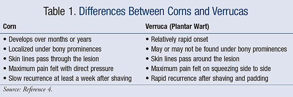
Differential Diagnosis: Verrucas (plantar warts) are warts that develop on the soles of the feet. They may sometimes be confused with corns and may need to be further investigated to reach a differential diagnosis ( TABLE 1). In verrucas, removal of the corneal layer of the skin reveals end arteries that may bleed or present as black dots if they are thrombosed.4

Management
The main goals for management of corns and calluses are to4:
- Provide symptomatic relief
- Determine mechanical etiology
- Formulate a conservative plan by advising on footwear and prescribing orthoses
- C onsider surgery if conservative measures fail.
Symptomatic Relief: Symptomatic relief of callosities can be provided through debridement, enucleation, keratolytic therapy, injection therapy, and orthotic therapy.
Patients should be instructed to soak the foot in warm water and use a pumice stone to remove the hard skin regularly.4 The skin may also be softened with emollient if necessary. Silicone sleeves provide good pain relief by cushioning and by slow release of mineral oil to soften the keratotic lesion.4
Pain associated with a callosity can be relieved to a certain extent by sharp debridement to reduce the amount of hyperkeratotic tissue.1 ,4 A scalpel can be used to remove the central keratin plug, with the administration of a local anesthetic if necessary. This provides complete pain relief. Recurrence can be prevented with gentle trimming every week and using a pumice stone or emery board after soaking the lesion in warm water for 20 minutes.4
Debridement, an enucleation of keratotic lesions, only provides short-term relief of symptoms unless the underlying cause is removed.
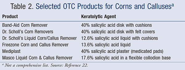
Since 1782, when Laforest used a concoction containing pork fat and the “mousse that forms around boats” as keratolytic therapy, an interesting variety of ointments, tinctures, and poultices have been proposed.19 Presently, topical agents containing salicylic acid, silver nitrate, and silicone and hydrocolloid wound dressings are used.20 Keratolytic agents such as salicylic acid can be applied directly as a solution or a pad. It has been shown that corn plasters containing 40% salicylic acid are more effective that nonmedicated placebo pads.21 They are available both as OTC and prescription products. Keratolytic agents must be used with caution, as overapplication can cause chemical burns.4 These agents should be avoided in neuropathic and immunocompromised patients.4 TABLE 2 lists some of the nonprescription products that are available to treat corns and calluses.22

More recently, hydrocolloid dressings that have a hydrating effect on the skin have been evaluated as a potential for the treatment of keratotic lesions. Preparations of sodium chloride, alcohol, and liquid silicone have also been tested for intralesional or sublesional injection.4 ,23 Deflective padding or padding with lamb’s wool can induce healing of maceration in painful interdigital soft corns.1
Footwear: Many lesions can be managed with the use of appropriate footwear. Patients should be advised to avoid tight shoes and chose styles that are low-heeled with a soft upper and a roomy toe box.24 Additionally, persons with corns or deformed toes should wear shoes with extra depth. Those with corns on the lateral aspect of the fifth toe and interdigital soft corns should wear shoes with extra width.4 In some cases, a cobbler may be able to stretch a shoe to relieve mechanical pressure on a lesion, or an orthotist may modify a shoe (e.g., add a medical wedge for a cavovarus foot) .4
On the other hand, loose shoes such as unlaced sneakers or open-back sandals may induce shearing forces on the edges of the weight-bearing area of the sole of the foot, resulting in marginal callus or heel fissures.4
Orthoses : Orthotic devices are often prescribed to redistribute mechanical forces in the foot and allow a lesion to heal.4 There are various types of orthoses, including doughnut-shaped corn pads, heloma shields, and silicone toe splints that relieve pressure from the tender central core in corns. In addition, silicone sleeves release mineral oil, thereby softening the lesion.4 Interdigital wedges made of Plastazote (a foam padding) or orthodigital splints made of silicone promote healing of an interdigital soft corn.4
Metatarsal pads placed proximal to the metatarsal head reduce the pressure of a metatarsal head on the underlying skin and are useful in the management of plantar calluses caused by weight-bearing stresses on the metatarsal heads. An adhesive felt pad may be used to transfer weight away from the painful area. This should be carefully cut, taking into consideration the size and the shape of the metatarsal heads. Specifically, the anterior edge of the metatarsal pad must be the full width of the metatarsal heads and narrow along the medial and lateral borders. Small, semicircular cuts that are large enough to accommodate the metatarsal heads are made into the distal edge of the pad. Further cuts that accommodate any one metatarsal head or any combination of metatarsal heads may be made.25
A ready-made, full-length shoe inlay made of padded leather or Plastazote may provide relief and can be moved from shoe to shoe. A customized shoe inlay of vacuum-molded Plastazote with added metatarsal relief is best at relieving pressure but can only be worn in extra depth shoes and not in most dress shoes.4
Surgical Procedures: A number of surgical options are available for those patients in whom conservative measures have not worked. Surgical procedures may be employed to remove bony prominences; change the mechanics of the foot; correct a claw toe, hammer toe, or mallet toe; or resect the prominent condyles to treat soft interdigital corns.26 It is useful to note that conservative treatment remains the best way of managing calluses, since metatarsal osteotomies produce unpredictable results and might result in the callus transferring to the adjacent metatarsal head.27 ,28
Conclusion
Corns and calluses are caused by a combination of inappropriate shoes, abnormal foot mechanics, and high levels of activity . Pharmacists can advise patients on correct footwear and the use of orthoses, keratolytic products, and regular debridement. In rare cases, surgery may be required to correct abnormal mechanical stresses.
REFERENCES
1. Freeman DB.
Corns and calluses resulting from mechanical hyperkeratosis.
Am
Fam Physician. 2002
;65:2277-2280.
2.
Menz HB, Lord SR. Foot pain impairs balance and functional ability in community dwelling older people. J Am Podiatric Med Assoc. 2001
;91:222-229.
3. Murray HJ, Young MJ, Hollis, et al. The association between
callus formation, high pressures and neuropathy in diabetic foot ulceration. Diabetic Med. 1996
;13:979-982.
4. Singh D, Bentley G, Trevino S.
Keratotic lesions, corns and calluses. BMJ. 1996
;312:1403-1406.
5. Day RD,
Reyzelman AM,
Harkless LB. Evaluation and management of the
interdigital corn: a literature review.
Clin
Podiatr Med Surg. 1996
;13:201-206.
6.
Haboush EJ, Martin RV.
Painful
interdigital
clavus (soft corn).
Treatment by skin plastic operation. J Bone Joint Surg. 1947
;29:756-757.
7. Gillett HG.
Interdigital
clavus: predisposition is the key factor of soft corns.
Clin
Orthopaedics Related Res. 1979
;142:103-109.
8.
Springett KP, Whiting MF, Marriott C. Epidemiology of plantar forefoot corns and calluses and the influence of the dominant side. The Foot. 2003
;13:5-9.
9. Merriman L, Griffiths C,
Tollafield D. Plantar lesion patterns.
Chiropodist (
Lond). 1987
;42:145-148.
10. George DH.
Management of
hyperkeratotic lesions in the elderly patient.
Clin
Podiatr Med Surg. 1993
;10:69-77.
11. Rubin L. Hyperkeratosis in response to mechanical irritation.
J Investigative
Dermatol. 1949
;13:313-315.
12. Thomas SE, Dykes PJ, Marks R. Plantar hyperkeratosis: a study of callosities and normal plantar skin.
J Investigative
Dermatol. 1985
;85:394-397.
13.
Silfverskiold JP.
Common foot problems. Relieving the pain of bunions,
keratoses, corns, and calluses. Postgrad Med. 1991
;89:183-188.
14.
Bonavilla EJ.
Histopathology of the
heloma durum; some significant features and their implications. J Am
Podiatr Assoc. 1968
;58:423-427.
15. Reed JF.
An audit of lower extremity complications in octogenarian patients with diabetes mellitus.
Int
J Low
Extrem Wounds. 2004
;3:161-164.
16. Williams AE, Bowden AP. Meeting the challenge for foot health in rheumatic diseases. The Foot. 2004
;14:154-158.
17. Jordan E,
Lekkas C,
Roscioli D, et al. Podiatric problems on a stroke rehabilitation unit.
Australian J Ageing. 1997
;16:222-224.
18. Sari-
Kouzel H, Hutchinson C, Middleton A, et al. Foot problems in patients with systemic sclerosis. Rheumatology. 2001
;40:410-413.
19.
Springett K, Parsons S, Young M, et al. The effect and safety of three
corn care products.
Brit J
Podiatr. 2002
;5:82-86.
20. Potter J. The use of salicylic acid in the treatment of dorsal corn and callus.
Brit J
Podiatr. 2000
;3:51-55.
21. Lang L, West S, Day S,
Simmonite N. Salicylic acid in the treatment of corns. The Foot. 1994
;6:145-150.
22. Pray WS. Chapter 8. Foot problems. In: Nonprescription Product Therapeutics. 2nd ed. Baltimore, MD: Lippincott Williams & Wilkins; 2006.
23.
Menz HB. Foot Problems in Older People: Assessment and Management. Philadelphia, PA: Churchill Livingstone; 2008.
24. Richards RN.
Calluses, corns, and shoes.
Semin
Dermatol. 1991
;10:112-114.
25. White SC. Padding and taping. In:
Valmassy RL, ed. Clinical Biomechanics of the Lower Extremities. St. Louis, MO: Mosby; 1996:368-389.
26. Coughlin MJ, Mann RA.
Lesser toe deformities. In: Mann RA, Coughlin MJ,
eds.
Surgery of the Foot and Ankle. 6th ed. St Louis, MO: CV Mosby; 1993:413-465.
27. Jimenez AL,
McGlamry ED, Green DR. Lesser ray deformities. In:
McGlamry ED,
McGlamry R,
eds.
Comprehensive Textbook of Foot Surgery. Baltimore, MD: Williams & Wilkins; 1987:57-111.
28.
Pontious J, Lane GD, Moritz JC, Martin W. Lesser metatarsal V-osteotomy for chronic intractable plantar keratosis.
Retrospective analysis of 40 procedures. J Am
Podiatr Med Assoc. 1998
;88:323-331.


