As the population of the United States ages and the baby boomers surge into the health care marketplace, pharmacists increasingly will be approached by patients with questions regarding common skin growths. The cause of common noncancerous growths is unclear, and even though enlargement may occur over time, most growths are benign. However, while most benign skin lesions do not invade other tissues and spread to other parts of the body—i.e., metastasize—a critical caveat is that they all must be watched by the patient and examined by a clinician if any changes occur.1
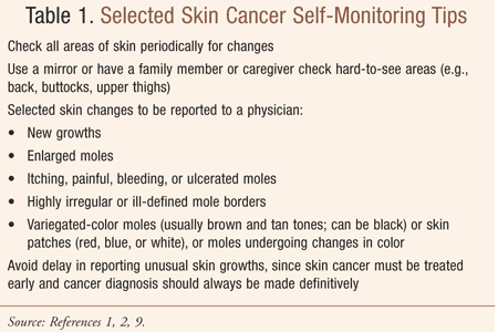
Skin cancer, while generally occurring on skin that has been most exposed to the sun, can in fact develop anywhere on the body. Therefore, skin self-monitoring (TABLE 1) and skin surveillance by a trained clinician are integral components of skin cancer identification and prevention. These monitoring measures are considered health care imperatives because skin cancers must be treated early and proper diagnosis of unusual skin growths should always be made definitively and without undue delay. Pharmacists are involved in this process through education, referral for evaluation and treatment, and counseling regarding appropriate medication management and follow-up of skin growths.

Additionally, although common skin growths are usually benign, they may be unacceptable to today’s seniors. Rather than tolerate unsightly or physically uncomfortable skin discolorations or growths, many older adults opt to seek medical attention to minimize the appearance of growths or eliminate them through medical, surgical, or pharmaceutical intervention, or a combination of these methods. In this regard, patients may find that because some procedures for removal of benign skin growths are considered cosmetic in nature, they may not be covered by the their health insurance plan.
Age-Related Changes of the Skin
Skin becomes drier, thinner, less elastic, and finely wrinkled with normal aging. Sun exposure over the years, however, is responsible for wrinkled, rough, and blotchy skin.2 While the appearance of a person’s skin changes with age, the most unwanted changes are due to chronic sun damage. People who have avoided the sun or blocked their skin from sun exposure over their lifetime often appear younger than their chronological age.
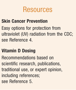
Importantly, the number of pigment-containing cells in skin, called melanocytes, decreases with age, so that older adults have less protection against ultraviolet (UV) radiation than their younger counterparts.2,3 Accordingly, use of sunscreen is important, since UV rays can damage skin in as little as 15 minutes.4 Patients should be guided regarding the proper type and use of sunscreen, and the FDA’s significant changes to sunscreen product labels (see RESOURCES).

Additionally, Vitamin D3 is synthesized in the skin when it is exposed to UVB rays from sunlight.5 Since aging skin functions less well and becomes less able to convert vitamin D to its active form, vitamin D supplementation with calcium for bone health in seniors should be considered (see RESOURCES).
Elevations and Discolorations of the Skin
Age spots (liver spots, lentigos): With an appearance similar to a freckle, an age spot is flat and light brown in hue; it is common after age 40 years.2,6 These skin discolorations develop after many years of sun exposure, and although they are referred to as liver spots, they have no relationship to the liver or liver function.5 Age spots appear most often on areas of skin likely to get UV exposure such as the face, shoulders, forearms, and back of the hands.2,6 Topical skin bleaching preparations, such as hydroquinone, may be used to lighten the pigmentation; patients should be counseled regarding burning, blistering, fissuring, or other skin reactions that may occur with hydroquinone use.6,7 Cryotherapy or laser treatment can be used to eliminate age spots.
Moles (nevi) and atypical moles (dysplastic nevi): A mole (nevus) is a flat or raised discoloration, usually dark in color, that can appear anywhere on the body. Once a mole emerges and develops, it may remain for decades; moles typically become more raised and less pigmented over the years.2,8 The removal of moles for cosmetic purposes is achieved by shaving or excision. A hairy mole should be adequately excised (versus being shaved) if the patient is concerned about hair growth; otherwise, hair regrowth will occur.8 Histologic examination should be performed on all moles that are removed. While the majority of moles are benign, some become cancerous. The presence of more than 20 moles indicates a higher-than-average risk for melanoma.8
Atypical moles are nevi with a slightly different clinical and histologic appearance. They are typically larger than other nevi (>6 mm in diameter) and primarily round (unlike many melanomas); however, they appear with indistinct borders and mild asymmetry.8 Melanomas, in contrast, have greater irregularity of color; in addition to being tan and brown, they can appear dark brown, black, red, and blue or with whitish areas of depigmentation.8 Patients with atypical moles are at increased risk of melanoma, with risk increasing as the number of atypical moles and level of sun exposure increase.8 While atypical moles may appear anywhere, they are more commonly found on covered areas such as the buttocks, breasts, and scalp. An individual may have the propensity to develop atypical moles based on heredity; or, moles may be sporadic, without apparent familial association.8 The presence of multiple atypical moles and melanoma in more than two first-degree relatives is referred to as familial atypical mole–melanoma syndrome, in which patients are at markedly increased risk (25 times) for melanoma.8 Self-monitoring for warning signs (TABLE 1) and professional skin surveillance are critical.
Seborrheic keratoses: These pigmented superficial epithelial lesions are generally referred to as seborrheic warts since the plaques are soft, friable, and wartlike in appearance; they can also appear as smooth papules. Typically, seborrheic keratoses present on the trunk or temples in mid to later life and vary in size. When lesions (1-3 mm) are noted on the cheekbones in black and Asian individuals, the condition is referred to as dermatosis papulosa nigra.8 Round or oval in shape and flesh-toned, brown, or black in color, these lesions have a characteristic “stuck on” appearance with a surface that is crusty, verrucous, velvety, waxy, or scaly. They are diagnosed clinically, grow slowly, are not premalignant, and do not require treatment. Removal may be sought if they rub against clothing, or become irritated, itchy, or cosmetically annoying; removal is achieved by cryotherapy (freezing with liquid nitrogen) or electrodesiccation and curettage following a local lidocaine injection.8
Acrochordons (skin tags): These pigmented or hyperpigmented, pedunculated (attached by a stalk) lesions usually appear as multiples on the neck, axilla, and groin. They are flesh-colored or more deeply pigmented and are small and soft, thus referred to as soft fibromas. While the lesions are usually asymptomatic, removal may be sought if they become irritating—causing a pinching or pulling sensation—or cosmetically unsightly. Removal entails cryotherapy, light electrodesiccation, or excision with a scalpel or scissors; histologic examination of all skin tags is the standard of care.9
Cherry angiomas: Most common after age 30, these bright cherry-red or purple benign skin elevations or spots are formed from overgrowths of blood vessels.2,10 While their cause is unknown, they tend to be inherited. These lesions can occur almost anywhere on the body; however, they typically appear on the trunk.10 Cherry angiomas vary in size, and while usually no larger than one-eigth inch (3 mm) in diameter, can be as large as approximately one-quarter of an inch.2,10 Although these benign growths usually do not require treatment, if they bleed often or affect appearance they may be removed by electrosurgery/cautery, cryotherapy, laser, or shave excision.10
Of note, with regard to the conditions listed above, dermal procedures may vary based on the extent of the condition, the clinician’s preference, or other considerations. Patient counseling by the pharmacist may include not only guidance regarding medication therapy for treatment (e.g., hydroquinone), but also preprocedure topical anesthetics (e.g., a topical cream combining lidocaine 2.5% and prilocaine 2.5%; a topical patch combining lidocaine 70 mg and tetracaine 70 mg) for local dermal analgesia.
Conclusion
While most noncancerous skin lesions do not invade other tissues and spread to other parts of the body, it is critical that all benign lesions be self-monitored in addition to being examined by a clinician if any changes occur. Pharmacist guidance regarding medication management in the treatment of benign skin growths and cancer prevention interventions is essential.
REFERENCES
1. Henry GI. Benign skin lesions. Overview of benign skin lesions. Medscape Reference. Drugs, Diseases and Procedures. Updated October 12, 2012. http://emedicine.medscape.com/article/1294801-overview. Accessed May 15, 2013.
2. Beers MH, Jones TV, Berkwits M, et al, eds. The Merck Manual of Health & Aging. Whitehouse Station, NJ: Merck Research Laboratories; 2004:11-12,430-435.
3. Aging changes in skin. MedlinePlus. Updated September 4, 2012. www.nlm.nih.gov/medlineplus/ency/article/004014.htm. Accessed May 15, 2013.
4. Skin cancer. Prevention. Centers for Disease Control and Prevention. Updated April 23, 2013. www.cdc.gov/cancer/skin/basic_info/prevention.htm. Accessed May 14, 2013.
5. Vitamin D. Dosing. MayoClinic.com. Updated September 1, 2012. www.mayoclinic.com/health/vitamin-d/NS_patient-vitamind/DSECTION=dosing. Accessed May 15, 2013.
6. Liver spots. MedlinePlus. Updated November 20, 2012. www.nlm.nih.gov/medlineplus/ency/article/001141.htm. Accessed May 15, 2013.
7. Epocrates, Version 4.5. Epocrates, Inc. www.epocrates.com. Accessed May 20, 2013.
8. Benign skin tumors. Moles, seborrheic keratoses. The Merck Manual. Revised September 2008; updated February 2012. www.merckmanuals.com/professional/dermatologic_disorders/benign_skin_tumors/introduction.html. Accessed May 3, 2013.
9. Benign skin tumors. Skin tags. The Merck Manual. Revised September 2008; updated June 2010. www.merckmanuals.com/professional/dermatologic_disorders/benign_skin_tumors/introduction.html. Accessed May 3, 2013.
10. Cherry angioma. MedlinePlus. Updated November 20, 2012. www.nlm.nih.gov/medlineplus/ency/article/001441.htm. Accessed May 15, 2013.
To comment on this article, contact rdavidson@uspharmacist.com.


