US Pharm. 2011;36(5):28-34.
One of the most feared sports and work injuries is a tear of the anterior cruciate ligament (ACL), which has ended or derailed the careers of numerous high-profile athletes. A torn ACL is very painful and can debilitate a person for several months and perhaps for life, although recovery for some is possible. In his book, professional football player Drew Brees describes “blowing out” his knee as a high school quarterback in 1995.1 However, following knee reconstruction surgery and rehabilitation, he went on to play for Purdue University and later the New Orleans Saints, who won Super Bowl XLIV in 2010. Other high-profile athletes who have sustained ACL tears include football quarterback Tom Brady, golfer Tiger Woods, and soccer player Frankie Hejduk.2
An ACL tear is one of the most common sports-related injuries, and its rehabilitative course may last 6 to 9 months.3 ACL tears may also occur during rough play, motor vehicle collisions, falls, and work-related injuries. About 80% of sports-related ACL tears are noncontact injuries.2 Most often, ACL tears occur when an athlete pivots or lands from a jump. The knee “gives out” from under the athlete when the ACL is torn. In addition to the ACL, other ligaments and the meniscus cartilage may also be damaged.
With greater emphasis on athletes making spectacular moves and dazzling plays, more stress is being placed on muscles and joints, possibly exceeding human limitations. In 2003, there were approximately 20 million visits to physicians’ offices because of knee injuries.3,4 Such injury is the most common reason reported for visiting an orthopedic surgeon. No other joint in the human body is as large or as important for smooth, powerful, and elegant movement than the knee. With every running stride or landing from a jump, the athlete’s knee absorbs and diffuses forces equivalent to eight to 10 times the body’s weight.5 It coils when pivoting, allowing the foot to point in one direction and the trunk in another. The knee is a weight-bearing joint that straightens, bends, twists, and rotates. All of these movements increase one’s risk of acute or chronic knee injuries.5-7
Female athletes are known to have a higher risk of injuring their ACL while participating in competitive sports.5 As more and more women participate in sports, the incidence of ACL injuries is increasing. Rates of ACL tears are about eight times more frequent in women than in men.7 Unfortunately, it is unclear as to why women are more susceptible to ACL injury. Some theories include8-10:
- Anatomic differences: These involve pelvis width, Q-angle, size of the ACL, and size of the intercondylar notch (where the ACL crosses the knee joint).
- Hormonal differences: The ACL has hormonal receptors for estrogen and progesterone, and it is believed that fluctuating hormone concentrations may play a role in ACL injuries, especially during different phases of the menstrual cycle.
- Biomechanical differences: Stability of the knee is dependent on static stabilizers (i.e., the ACL and other major knee ligaments) and dynamic stabilizers (i.e., muscles and tendons) that surround the knee joint. Some people believe that women land differently when jumping, possibly because of certain physical activities when they were young. Women are now known to have different biomechanical movements of the knee joint when pivoting, jumping, and landing—activities that often lead to an ACL injury.
Knee Anatomy
The knee performs multiple tasks perfectly because just below the skin lies a latticework of ligaments and tendons that hold together multiple muscles and bones. The knee is made up of three bones: the lower end of the thigh bone (femur); the end of the shin bone (tibia); and the knee cap (patella). Articular cartilage is a type of slick, hard, bonelike, flexible, white connective tissue that covers the surface ends of the tibia and femur to reduce friction and allow bones to move easily against each other. Another important structure in the knee—the meniscus—is a soft, tough, rubbery crescent-shaped wedge similar to a shock absorber between the femur and the tibia. The meniscus helps distribute the weight of the body across the knee joint, lubricate and protect the articular cartilage from wear and tear, stabilize the knee when one slides and turns, and limit extreme knee flexion and extension. There are two types of meniscus: the lateral meniscus, which slides forward and backward, and the medial meniscus, which helps the ACL and medial collateral ligament (MCL) stabilize the knee, absorbing up to 50% of the load applied to the inside of the knee.5-7
Just behind the knee cap are four essential ligaments that connect the femur and the tibia: 1) the MCL, which runs along the inside of the knee joint and provides stability to the medial (inner) part of the knee; 2) the lateral collateral ligament (LCL), which runs along the outside of the knee joint and provides stability to the lateral (outer) part of the knee; 3) the ACL, which is in the center of the knee and limits rotation and forward leg movements; and 4) the posterior cruciate ligament (PCL), which is in the center of the knee and limits backward leg movements.5-7
Knee Joint Injuries
Knee pain is commonly caused by trying to do “too much too soon” when one has not exercised for a long period of time—especially high-impact activities; e.g., aerobics, brisk walking, running or jumping on hard surfaces or uneven ground, and excessive running up and down stairs. When muscles and tendons are stressed even slightly beyond their capabilities, microscopic tears can occur. These tears must be given a chance to heal before being subjected to the same or vigorous activity to avoid overuse injury.5-7
Ligament Tear Injuries: These are common injuries among active individuals and athletes. Of the four major ligaments found in the knee, the ACL and MCL are most often injured in sports and/or physical stress injuries. PCL and LCL ligament injuries occur less often. In general, twisting the knee or falling often causes torn ligaments. Individuals at high risk are those participating in sports that include running, jumping, sudden stopping, or abrupt changes of direction, such as soccer, basketball, volleyball, tennis, and baseball, as well as contact sports like football, wrestling, and hockey.5-7
ACL Injury—“Blowing Out Your Knee”: Injuries to the ACL are often sports related. However a torn, stretched, or ruptured ACL can also be caused by repetitive physical stress, such as excessive pivoting or twisting of the knee. ACL injuries generally cause swelling, stiffness, and pain. Many times, a “popping” noise can be heard at the time the injury occurs. This injury can occur as a result of changing direction rapidly, slowing down abruptly when running, and landing forcefully from a jump so as to cause tears in the ACL. Male and female athletes who participate in skiing or basketball or who wear cleats, such as football players, are very susceptible to ACL injuries.5-7
When the ACL is torn and the signature loud “pop” is heard, intense pain follows and, within an hour, swelling occurs. Moderate-to-severe pain is very common. Initially, the pain is sharp and then becomes more of an ache or throbbing sensation as the knee swells. Since the ACL is the major knee stabilizer, an injury to it will cause the knee to give out or buckle when a person tries to walk or change direction. A common test of a suspected ACL tear is to bend the knee and see if the ligament can prevent the tibia from moving forward on the femur.5-7
MCL Injury: Injuries to the MCL are usually caused by a severe, direct blow to the outside of the knee. These types of injuries often occur in contact sports (e.g., football, soccer) and in skiing.5-7
PCL Injury: These injuries may occur when an athlete receives a hard blow to the front of the knee or makes a simple misstep on the playing field.5-7
Overview of Pain
Pain is defined as an unpleasant sensory and emotional experience associated with actual or potential tissue damage.11 The peripheral nervous system and the central nervous system (CNS) constitute an integrated system that provides a pathway for pain transmission. Transduction is the term used to describe the phenomena associated with the initiation of the pain signal. Pain receptors are found on the peripheral end plates of afferent neurons. The sensation of peripheral pain begins in afferent (conducts nerve impulses from the periphery to the brain) neurons called nociceptors.12 There are two types of these neurons and they may be stimulated by mechanical, thermal, hormonal, chemical, and serotonin stimuli. Delta fibers transmit fast-traveling, myelinated stimuli and sense sharp, stinging, cutting, or pinching pain. C fibers transmit slow-traveling, unmyelinated stimuli and sense dull, burning, or aching pain. Of the five opiate receptors, only three are activated with narcotic analgesics: Mu activation results in supraspinal analgesia, respiratory and physical depression, euphoria, sedation, reduced gastric motility, and miosis; kappa activation results in spinal analgesia and less respiratory depression, miosis, sedation, and gastric motility than mu; and delta activation results in analgesia, dysphoria, and hallucinations.12

Treatment of Knee Injuries and Pain Management
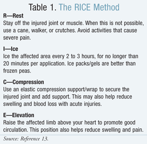
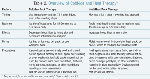
Treatment regimens for knee injuries must be individualized to the type and severity of injury and often involve a combination of different treatment methods, including cold and heat therapy (TABLES 1 and 2). Typical treatment regimens for knee injuries often involve the RICE (Rest, Ice, Compression, Elevation) method (TABLE 1).13 RICE is an effective way to treat acute knee injuries. This method is first-line therapy and should be implemented within the first 72 hours of injury or upon occurrence of sore muscles, joint ache, or swelling.

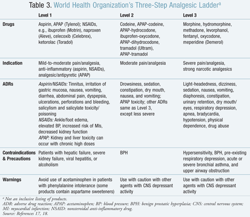
Treatment Options Using Oral Medication: Pharmacologic management for acute pain includes nonopioid and opioid medication.14 Nonopioid medications include nonsteroidal anti-inflammatory drugs (NSAIDs) available on a prescription and nonprescription basis
(TABLE 3). NSAIDs produce pain relief by inhibition of cyclooxygenase (COX-1 and COX-2), which catalyzes the production of proinflammatory prostaglandins from arachidonic acid. Prostaglandins mediate inflammation and increase pain sensations and swelling through inflammatory mechanisms.15 Opioid medications mimic the actions of endorphins by interacting primarily with mu opioid receptors but also with kappa and delta receptors in the brain, and opioid receptors in ascending pathways may work to inhibit nociceptive signals.12
Orthopedic Physician Evaluation and ACL Reconstruction Surgery: ACL reconstruction surgery is the standard treatment for adults and some young, active people who sustain an ACL tear. But what happens when that person is a child? Should ACL surgery be delayed until the child is older, or should ACL reconstruction be performed before skeletal maturity? The concern of performing ACL surgery in children is the risk of causing a growth disturbance. Growth plate problems as a result of ACL surgery could potentially lead to early growth plate closure or alignment deformities.3,4
Rehabilitation Therapy: To repair ACL injuries in an active athlete, ACL reconstruction surgery and a lengthy rehabilitation period of 6 to 9 months (e.g., physical therapy and/or occupational therapy following surgery) are often required.3
What Can Be Done to Prevent ACL Injuries?
The best way to reduce the risk of ACL injury is with the use of neuromuscular training programs before engaging in sports activities. Neuromuscular training is the process of teaching your body better biomechanic movements and improved control of dynamic stabilizers. Neuromuscular programs have been designed to address deficits in dynamic stabilization of the knee. Several programs have been designed, most of which involve stretching, plyometrics, and strengthening. These programs teach the athlete how to land from a jumping position, to pivot side-to-side, and to move the knee without placing as much force on the ACL.
One such program is the PEP program developed at the Santa Monica Orthopaedic and Sports Medicine Research Foundation.9,16 The PEP of the program’s title stands for prevent injury; enhance performance. Components of the program include:
• Warm-up: Warming up muscles and cooling down greatly reduce risk of injury. Moves include line-to-line jogging, shuttle running, and backward running.
• Stretching muscles (calf, quadriceps, hamstring, and inner thigh): Never stretch cold muscles.
• Hip flexor stretch: Elongate the hip flexors of the front of the thigh.
• Strengthening: Increase leg strength by doing walking lunges, Russian hamstring exercises, and single-toe raises.
• Plyometrics: Help build power, strength, and speed by doing, e.g., lateral hops over cones, forward/backward hops over cones, single leg hops over cones, vertical jumps with headers, and scissors jumps.
• Agilities: Shuttle run with forward/backward running, diagonal runs, and bounding runs.
• Alternative exercises (warm down and cool down): Bridging with alternating hip flexion, abdominal crunches, single and double knee to chest (supine), figure-four piriformis stretches, and seated butterfly stretches.

Role of the Pharmacist in the Prevention of Pain Medication Addiction
Following a knee injury or surgery, pain management may include prescription nonnarcotic (TABLE 3, Level 1) or narcotic (TABLE 3, Level 2 or 3) medication. Depending on the surgeon and degree of knee damage/repair, in addition to the RICE regimen, he or she may initially prescribe narcotic medication to control severe pain short-term and then switch to nonnarcotic medication for long-term pain management. Cold/ice and heat therapy seem to be the mainstay of joint and muscle pain management. NSAIDs help relieve inflammation and thus address the problem that causes the swelling and pain. Narcotics, on the other hand, relieve pain very well but do not diminish the problem causing the pain, but rather cover up the painful symptoms.
Patients prescribed narcotics may be worried about addiction. Unfortunately, some patients are so fearful of narcotic pain medication that they may not take the drug and may be unable to perform their proper rehabilitation. Narcotic medications are best used for short-term control of acute pain (immediately following injury or surgery). Addiction to narcotics may occur with longer-term use and when patients take medication for the effects rather than for pain relief. Patients with a history of substance abuse may require careful monitoring for compliance issues. They may present with various reasons to get early, inappropriate refills on a previously filled narcotic prescription, or try to fill another new narcotic pain prescription from a different physician. Hopefully, this information will assist pharmacists in effectively helping patients avoid drug diversion or dependency and in dealing better with noncompliant patients (i.e., those taking too much, too often) before they become addicted.
REFERENCES
1. Brees D, Fabry C. Coming Back Stronger: Unleashing the Hidden Power of Adversity. Carol Stream, IL: Tyndale House Publishers, Inc; 2010.
2. Cluett J. What you need to know about ACL tears. About.com Guide. http://orthopedics.about.com/
3. McCarty LP III, Bach BR Jr. Rehabilitation after patellar tendon autograft anterior cruciate ligament reconstruction. Techniques Orthop. 2005;20:439-451.
4. Common knee injuries. American Academy of Orthopaedic Surgeons. http://orthoinfo.aaos.org/
5. Reynolds G. The complete guide to your knee. Men’s J. 2010;18:53-63.
6. Koopman WJ, Boulware DW, Heudebert G, eds. Clinical Primer of Rheumatology. Philadelphia, PA: Lippincott Williams & Wilkins; 2003.
7. Ruddy S, Firestein GS, Budd RC, et al, eds. Kelley’s Textbook of Rheumatology. Philadelphia, PA: WB Saunders Co; 2008.
8. Slauterbeck J, Arendt E, Beynnon BD, et al. ACL injuries in women: why the gender disparity and how do we reduce it? Orthop Today. 2003;23:1. www.orthosupersite.com/view.
9. Griffin LY, Agel J, Albohm MJ, et al. Noncontact anterior cruciate ligament injuries: risk factors and prevention strategies. J Am Acad Ortho Surg. 2000;8:141-150.
10. Hewett TE, Lindenfeld TN, Riccobene JV, Noyes FR. The effect of neuromuscular training on the incidence of knee injury in female athletes: a prospective study. Am J Sports Med. 1999;27:699-706.
11. Merskey H, Bogduk N, eds. Part III: Pain terms, current list with definitions and notes on usage. In: Classification of Chronic Pain. 2nd ed. Seattle, WA: IASP Press; 2004:209-214. www.iasp-pain.org. Accessed June 23, 2010.
12. Vanderah TW. Pathophysiology of pain. Med Clin N Am. 2007;91:1-12.
13. Self-care of self-limited pain. In: Albrant DH, ed. The American Pharmaceutical Association Drug Treatment Protocols. 2nd ed. Washington, DC: American Pharmaceutical Association; 2001:424-425.
14. American Pain Foundation. Pain Resource Guide: Getting the Help You Need. 2007; revised 2009. www.painfoundation.org/learn/
15. Rao PN, Knaus EE. Evolution of nonsteroidal anti-inflammatory drugs (NSAIDs): cyclo-oxygenase (COX) inhibition and beyond. J Pharm Pharmaceut Sci. 2008;11:81s-110s.
16. Gilchrist J, Mandelbaum BR, Melancon H, et al. A randomized controlled trial to prevent noncontact anterior cruciate ligament injury in female collegiate soccer players. Am J Sports Med. 2008;36:1476-1483.
17. Kastrup EK. Drug Facts and Comparisons. St. Louis, MO: Facts & Comparisons; 2011.
18. World Health Organization. Cancer Pain Relief, With a Guide to Opioid Availability. 2nd ed. Geneva, Switzerland: World Health Organization; 1996.
To comment on this article, contact rdavidson@uspharmacist.com.


