US Pharm. 2010;35(6):38-44.
The lifetime incidence of developing a foot ulcer may be as high as 25% for the 24 million Americans with diabetes.1 Ulcers, defined as any breaks in the cutaneous barrier, are the most frequent type of diabetic foot wounds and usually extend through the dermis.2 Foot ulcers can cause substantial morbidity, arising from physical and emotional effects, loss of productivity, and financial expense, as well as increased mortality. Consequences could be dire; a foot ulcer precedes 85% of nontraumatic amputations, and mortality can range from 39% to 80% at 5 years postamputation.3,4
Background on Foot Ulcer Development
Poor metabolic control plays a role in foot ulcer development, underscoring the importance of reducing hemoglobin A1C (HbA1C) to the American Diabetes Association (ADA) guideline goal of less than 7%.5 Intensive control has reduced neuropathy by 57% in type 1 diabetic patients. Each 1% reduction in HbA1C has decreased microvascular complications by 25% in type 2 diabetes.6,7 Diabetic peripheral neuropathy (DPN) is a major microvascular complication that, along with excessive plantar pressure associated with deformity and trauma, can play a major role in ulcer development.
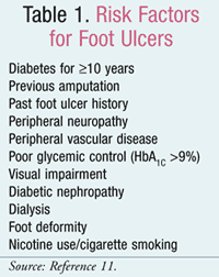
DPN can involve all three types of nerve dysfunction: sensory, autonomic, and motor. Sensory neuropathy results when the patient develops a lack of protective sensation. A painless thermal or mechanical injury occurs and remains undetected. Thus, the classic foot trauma case: A patient with diabetes unknowingly steps on a nail, and the foot becomes infected, which leads to serious complications. Autonomic dysfunction leads to reduced perspiration, which compromises skin integrity. Dry skin can easily develop cracks and fissures if not rehydrated properly, which leads to infections.8 Motor dysfunction develops in patients with foot deformities, such as bunions, hammer toes, and claw toes. Foot ulcers typically develop on forefoot plantar areas of maximum friction, such as the hallux and second metatarsophalangeal joint. However, altered gait resulting from foot deformities can lead to unusual points of friction and ulcer formation.1 A profound loss of protective sensation (LOPS), which increases the risk of ulceration by sevenfold, usually develops after DPN progression.9,10 The LOPS leads to erythema, blistering, callus formation, and, ultimately, ulceration. Patients with ulcers and peripheral vascular disease or prolonged wound healing are susceptible to adverse outcomes, and onychomycosis is more common. Risk factors for ulcer development and subsequent amputations can be found in TABLE 1.11
Assessment of the Diabetic Foot
The ADA recommends an annual, comprehensive foot examination consisting of a global inspection, assessment of pedal pulses, and testing for LOPS.5 General foot self-care education and screening for peripheral arterial disease are also recommended. Smokers, or those with LOPS, foot deformities, or a history of lower extremity complications should be referred to foot care specialists.5 The ADA recommends a multidisciplinary approach; thus, this article will emphasize pharmacy care components where applicable.
Key components of a comprehensive foot examination include a history, global inspection, and dermatologic, musculoskeletal, neurologic, and vascular assessment.12 Risk factors (TABLE 1) are assessed through the patient history. Previous foot ulcerations or amputations are especially high-risk conditions, and patients should be referred to a foot specialist. Peripheral vascular disease is twice as common in patients with diabetes, while cigarette smoking is a risk factor for neuropathy and vascular disease.13 A causal association has been shown between nicotine use and foot ulceration and amputation.1,14
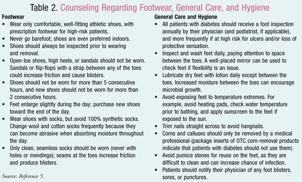
Global Inspection: Both the foot and footwear should be inspected. For example, shoes could have excessive wear, be too small or narrow, or the toe box could be too low. High-risk patients should purchase prescription shoes with ample padding and a deep, wide toe box.15 Shoe inserts can also equally distribute pressure. Some patients may need custom-molded shoes. Lower-risk patients can wear general athletic shoes if the shoes are well-fitting and of good quality. Counseling points specific to footwear are given in TABLE 2.
Dermatologic and Musculoskeletal Assessment: Any deformities or abnormal erythema, blisters, or calluses should be noted, including an inspection between the toes. Excessive dryness or swelling should be further evaluated. Nails should not be thickened, too long, ingrown, or infected. High temperature gradients between feet could predict ulceration or vascular disease.
Neurologic Assessment: Tests to identify LOPS are simple to perform and include: 1) pressure assessment with the nylon filament Semmes-Weinstein monofilament test; 2) vibration testing with a 128-Hz tuning fork; 3) testing for pinprick sensation; 4) ankle reflex assessment; and 5) vibration perception threshold testing with a biothesiometer.12 Each test has been evaluated in well-conducted prospective clinical studies and can identify LOPS.16 The monofilament test (the one most frequently used) plus any one of the other tests is recommended to rule out LOPS. An abnormal finding for either test would indicate LOPS.12
Clinically significant large-fiber neuropathy is identified through pressure assessment with the monofilament test and is conducted as follows: a 5.07 monofilament buckles when 10 g of force is applied perpendicular to the skin. Once buckling occurs within 1 second, the monofilament is released. The patient identifies with a “yes” or “no” and eyes closed whether the nylon probe is touching skin. The patient should be able to identify the spot of monofilament placement. Four to 10 sites should be tested on each foot. Testing on the dorsal surface of the great toe and at the base of the first, third, and fifth metatarsals can identify 90% of patients with LOPS. Testing up to 10 total sites, in no certain order, is also recommended.16 A “positive” or “negative” entered on the chart indicates sensation or a lack of sensation, respectively. Callused areas or scar tissue should be avoided. Any area with a lack of sensation may be retested; then the probe is inserted onto adjacent areas to map the extent of LOPS.16
A vibrating 128-Hz tuning fork is placed at the tip of the great toe of each foot. If the patient stops detecting vibration while the examiner can still sense it, this is an abnormal response. For the pinprick test, with the patient’s eyes closed, a pin is placed onto the dorsal surface of the great toe with enough force only to depress the skin. An abnormal test result would be inability of the patient to detect the depression.16
For the ankle reflex, the Achilles tendon is stretched until the ankle is in a neutral position. When a tendon hammer is struck, an abnormal result is total absence of reflex. The last test uses a biothesiometer, which is a handheld device that can assess the vibration perception threshold. The stylus is placed at the great toe, and the amplitude increases until the patient detects the vibration. This gives a quantitative measurement, and a vibratory perception threshold greater than 25 volts is an abnormal result.16
Vascular Assessment: A vascular assessment is not usually extensive.17 The easily palpated dorsalis pedis and posterior tibialis are the pedal pulses. The dorsalis pedis is at the top of each foot between the great toe and the one adjacent. The posterior tibialis is posterior to the ankle. The pulses can be “present” or “absent,” with the latter screened further with the ankle-brachial index (ABI).17
The ABI, which is the ratio of systolic blood pressure in the ankle to that of the brachial artery, further assesses for peripheral arterial disease (PAD). If the ABI is 0.9 or less, this suggests PAD, while greater than 0.9 would be a “normal” result. It should be measured annually for patients with diabetes over age 50 and considered for younger patients with multiple PAD risk factors.16
Treatment of Diabetic Ulcers
Because initial OTC treatment is common, the pharmacist should assess progression of healing. If the wound is not healing within 4 weeks, the pharmacist should refer the patient to a specialist. A wound unhealed after 4 weeks has worse outcomes, including amputation.17
Infection, though not always present, is indicated by purulence or at least two of the following: warmth, pain, tenderness, induration, erythema, or swelling.18 Cellulitis or signs of systemic response can also occur, including fever, sweats, chills, tachycardia, hypertension, and hyperglycemia. Thus, appropriate treatment depends on determining the severity of infection.
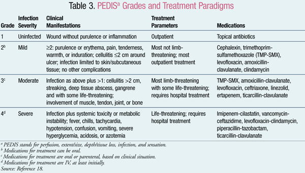
One evaluation system is summarized by the acronym PEDIS, which stands for perfusion, extent and size of the ulcer, depth or tissue loss, infection, and sensation.18 Categories based on infection severity range from grade 1 (“no infection”) to grade 4 (“severe”). While “mild” infections (grade 2) can be easily treated, “moderate” infections (grade 3) can be complicated and limb-threatening. TABLE 3 explains the classification system in further detail; however, presenting symptoms can be varied. In fact, more than half of patients with a limb-threatening infection do not have systemic symptoms.18 Thus, pharmacists should refer patients with grade 2 or above to a specialist. Even patients with an uninfected foot wound should be referred if healing has not progressed within 4 weeks.18
General treatment measures consist of debridement, wound culturing, off-loading, wound dressings, observation, control of hyperglycemia, and infection control. Health care specialists use a scalpel and forceps to debride necrotic tissue; this is essential for quick healing. The wound is swabbed and cultures analyzed while empiric therapy is begun.17
Off-loading is avoidance of all mechanical stress on the injured foot.17 This includes bed rest, surgical shoes, or foam dressings with crutches. Because patients frequently tire of off-loading, they must be counseled that healing will be unsuccessful if they constantly use their foot. In fact, off-loading should be continued until the wound is healed.
Although different types of dressings have been studied, no particular dressing has shown superiority. For example, antimicrobial dressings could be beneficial, but routine use cannot be recommended at this time.18 Observation should ensure that healing progresses, even for severe ulcers, over the first month.
Pharmacists should also emphasize the importance of metabolic control. While the ulcer heals, patients may experience more hyperglycemia than usual. Thus, diet and exercise should coincide with possible escalation of medication therapy. The effect of poor glycemic control on nephropathy, retinopathy, and neuropathy should be discussed, with frequent follow-up to monitor both blood glucose and infection control.
Infection Control
Patients commonly pursue OTC treatment at the pharmacy for an uninfected ulcer. Although PEDIS grade 1 ulcers do not require prescribed antibiotics, others require referral to a physician to evaluate if they are limb-threatening, life-threatening, or require hospitalization. Patients with severe infections (grade 4) need hospitalization, as these are life-threatening. Mild or moderate (PEDIS grades 2 and 3) infections may need brief hospitalization for observation or if complicating factors are present. However, most patients with mild or moderate infections can be treated on an outpatient basis.18
Initial empiric therapy is based on infection severity. Mildly infected wounds may only require topical antibiotics. A relatively narrow spectrum of agents may need to be covered (gram-positive cocci, the predominate organism) for mild-to-moderate infections. In contrast, severe infections should cover gram-negative rods and anaerobes. All severe and some moderate infections—essentially any that are limb-threatening—should at least be initially treated with IV antibiotics.18
Duration of therapy also depends on severity. Treatment for 1 to 2 weeks may be sufficient for mild infections, while moderate and severe should be treated for 2 to 4 weeks. Length of treatment also depends on effectiveness of concomitant therapy (e.g., debridement). Of interest, no single medication or combination has shown significant superiority.18 If the patient fails to respond to an initial antibiotic course, all antimicrobials should be discontinued and therapy directed at culture susceptibility. Possible choices of antibiotics for each level of severity can be found in TABLE 3.
The Pharmacist’s Role
Pharmacists are effective in the care of diabetes, including management of the diabetic foot. Optimal care begins with counseling, and TABLE 2 specifies discussion points to share with patients. In addition to counseling, pharmacists can have an impact by encouraging yearly exams to monitor for LOPS and by helping the patient gain metabolic control. Some pharmacists maintain foot care services for patients with diabetes, providing proper footwear and monitoring for problems. Pharmacists are especially skilled at treatment and infection control. It is also important for pharmacists to remember to refer patients with increasing ulcer severity, or a lack of progression of healing, for further assessment and evaluation. Pharmacists who adhere to these principles will be able to assist in the management of the diabetic foot.
REFERENCES
1. Singh N, Armstrong DG, Lipsky BA. Preventing foot ulcers in patients with diabetes. JAMA.
2005;293:217-228.
2. Lazarus GS, Cooper DM, Knighton DR, et al. Definitions and guidelines for assessment of wounds and evaluation of healing. Arch Dermatol. 1994;130:489-493.
3. Reiber GE. The epidemiology of diabetic foot problems. Diabet Med. 1996;13(suppl 1):S6-S11.
4. Reiber GE. Epidemiology of foot ulcers and amputations in the diabetic foot. In: Bowker JH, Pfeifer MA, eds. The Diabetic Foot. 6th ed. St. Louis, MO: Mosby; 2001:13-32.
5. American Diabetes Association. Standards of medical care in diabetes—2010. Diabetes Care. 2010;33(suppl 1):S11-S54.
6. The Diabetes Control and Complications Trial Research Group. The effect of intensive treatment of diabetes on the development and progression of long-term complications in insulin-dependent diabetes mellitus. N Engl J Med. 1993;329:977-986.
7. UK Prospective Diabetes Study (UKPDS) Group. Effect of intensive blood-glucose control with metformin on complications in overweight patients with type 2 diabetes (UKPDS 34). Lancet. 1998;352:854-865.
8. Kalish J, Hamdan A. Management of diabetic foot problems. J Vasc Surg. 2010;51:476-486.
9. Reiber GE, Vileikyte L, Boyko EJ. Causal pathways for incident lower-extremity ulcers in patients with diabetes from two settings. Diabetes Care. 1999;22:157-162.
10. Young MJ, Breddy JL, Veves A, Boulton AJ. The prediction of diabetic neuropathic foot ulceration using vibration perception thresholds. A prospective study. Diabetes Care. 1994;17:557-560.
11. Lavery LA, Armstrong DG, Vela SA, et al. Practical criteria for screening patients at high risk for diabetic foot ulceration. Arch Intern Med. 1998;158:157-162.
12. Boulton AJ, Armstrong DG, Albert SF, et al. Comprehensive foot examination and risk assessment: a report of the task force of the foot care interest group of the American Diabetes Association, with endorsement by the American Association of Clinical Endocrinologists. Diabetes Care. 2008;31:1679-1685.
13. Gregg EW, Sorlie P, Paulose-Ram R, et al. Prevalence of lower-extremity disease in the US adult population ≥40 years of age with and without diabetes: 1999-2000 National Health and Nutrition Examination Survey. Diabetes Care. 2004;27:1591-1597.
14. Boyko EJ, Ahroni JH, Stensel V, et al. A prospective study of risk factors for diabetic foot ulcer. The Seattle Diabetic Foot Study. Diabetes Care. 1999;22:1036-1042.
15. Tyrrell W. Orthotic intervention in patients with diabetic foot ulceration. J Wound Care. 1999;8:530-532.
16. Boulton AJ, Kirsner RS, Vileikyte L. Clinical practice. Neuropathic diabetic foot ulcers. N Engl J Med. 2004;351:48-55.
17. American Diabetes Association. Consensus Development Conference on Diabetic Foot Wound Care: 7-8 April 1999, Boston, Massachusetts. Diabetes Care. 1999;22:1354-1360.
18. Lipsky BA, Berendt AR, Deery HG, et al. Diagnosis and treatment of diabetic foot infections. Clin Infect Dis. 2004;39:885-910.
To comment on this article, contact rdavidson@uspharmacist.com.


