US Pharm. 2014;39(12):HS-27-HS-32.
Acute pancreatitis (AP) is sudden inflammation of the pancreas that may be mild or life-threatening. It may involve other regional tissues or remote organs. Severe abdominal pain is the predominant symptom, and blood tests and imaging tests help clinicians make the diagnosis. Whether mild or severe, acute pancreatitis usually requires hospitalization.1Acute pancreatitis affects men more often than women. The three most common causes of pancreatitis in the United States are heavy alcohol use, gallstones, and medications. The condition develops when the gallstones travel out of the gallbladder into the bile ducts, where they block the opening that drains the common bile duct and pancreatic duct. Genetics may be a factor in some cases. Sometimes, the cause is not known.1
The range of disease is from self-limiting to fatal, with an incidence and mortality rate that increases with age. Patients with inflammatory bowel disease (IBD) seem to be at increased risk for AP. In the elderly, gallstones result in a higher incidence of organ failure and death.2
Patients with AP usually seek urgent medical attention for the sudden onset of severe pain of the upper abdomen that radiates to the back. The onset of pain may be related to a recent rich, fatty meal or an alcohol binge.2
Clinical signs and symptoms, causes and etiology, diagnosis, complications, and therapy of acute pancreatitis are briefly discussed in this article.
Signs and Symptoms
Almost everyone with acute pancreatitis has severe and constant abdominal pain in the upper abdomen, below the sternum. The pain penetrates to the back in about 50% of people. With gallstones, the pain usually starts suddenly and reaches its maximum intensity in minutes. With alcoholism, pain develops over a few days. The pain remains steady and severe, with a penetrating quality, and persists for days. Some people have only slight abdominal tenderness, and in 5% to 10% of patients there is no pain at all.3
Coughing, vigorous movement, and deep breathing may worsen the pain. Sitting upright and leaning forward may provide some relief. Most people feel nauseated and have to vomit, sometimes to the point of not producing any vomit. Often, even large doses of an injected opioid analgesic do not completely relieve pain.3
Some people, especially those with alcohol abuse, may never develop any symptoms besides moderate pain. Conversely, others look sick, sweat, have a fast pulse (100-140 beats per minute) and shallow, rapid breathing. Rapid breathing is due to inflammation and the accumulation of fluid in the chest cavity.2,3
With acute pancreatitis, body temperature may be normal at first but will increase in a few hours to between 100°F and 101°F (37.7°C and 38.3°C). Blood pressure tends to fall when a person with AP stands, causing faintness. As the disease progresses, people tend to be less and less aware of their surroundings—some are nearly unconscious. Occasionally, the whites of the eyes (sclera) become yellowish. In certain people, the initial symptom may be shock or coma.1,3
Causes and Etiology
Normally, the pancreas secretes pancreatic fluid through the pancreatic duct to the duodenum. This pancreatic fluid contains inactive digestive enzymes and inhibitors that inactivate any enzymes that become activated on the way to the duodenum. Blockage of the pancreatic duct by a gallstone stuck in the sphincter of Oddi stops the flow of pancreatic fluid. Usually, the blockage is temporary and causes limited damage, which is soon repaired. But if the blockage remains, activated enzymes accumulate in the pancreas, overwhelm the inhibitors, and begin to digest the cells of the pancreas, causing severe inflammation.4
The pathogenesis of acute pancreatitis is not fully understood. Nevertheless, a number of conditions are known to induce this disorder with varying degrees of certainty, with gallstones and chronic alcohol abuse accounting for 75% of cases in the United States. The number of cases diagnosed as “idiopathic” will decrease as our understanding of the disease improves.4
Gallstone Pancreatitis: Because the gallbladder and pancreas share a drainage duct, gallstones that lodge in this duct can prevent the normal flow of pancreatic enzymes and trigger acute pancreatitis.
Alcoholic Pancreatitis: Alcohol is a common cause of acute pancreatitis. Alcoholic pancreatitis is more common in individuals who have a long history of alcohol abuse.
Drug-Induced Pancreatitis: A number of drugs used to treat medical conditions can trigger acute pancreatitis. Published case reports of drug-induced AP exist for at least 40 drugs of the top 200 most prescribed medications. The six drug classes commonly associated with AP are hydroxymethyl glutaryl coenzyme A reductase inhibitors, angiotensin-converting enzyme inhibitors, estrogens/hormone-replacement therapy, diuretics, highly active antiretroviral therapy, and valproic acid.5
Hereditary Conditions: Acute pancreatitis can be caused by hereditary conditions, such as familial hypertriglyceridemia and hereditary pancreatitis. These conditions usually occur in children and young adults.
Unexplained: No underlying cause can be identified in about 20% of people with acute pancreatitis. This condition is called idiopathic pancreatitis. Only a small proportion of this group will experience additional attacks over time.
ERCP-Induced Pancreatitis: Endoscopic retrograde cholangiopancreatography (ERCP) is a procedure that evaluates the gallbladder or pancreas. Acute pancreatitis develops in about 3% to 5% of people who undergo ERCP. Most cases of ERCP-induced pancreatitis are mild. This is a technique that uses x-ray to view the patient’s bile and pancreatic ducts.6
The functions of the common bile duct and the pancreatic duct are to drain the gallbladder, liver, and pancreas; the two main ducts convey the bile and the pancreatic juice through the papilla into the duodenum. The most common reason that someone would need an ERCP is a blockage of one of these ducts (often due to gallstones).3,6
Endoscopic techniques look for abnormalities such as blockages, tissue irregularities, problems with the flow of bile or pancreatic fluid, stones, or tumors, and endoscopic methods have replaced surgery in most patients with common bile duct and pancreatic disease.4
While any patients who need ERCP are hospitalized, the procedure is sometimes done on an outpatient basis.
Complications
Damage to the pancreas may permit activated enzymes and toxins—such as cytokines—to enter the bloodstream and cause low blood pressure and damage to organs outside of the abdominal cavity, such as the lungs and kidneys. The part of the pancreas that produces hormones, especially insulin, is usually not affected.6
About 20% of people with AP develop some swelling in the upper abdomen. This may occur because the stomach is distended or has been moved out of place by a mass in the pancreas that causes swelling, or because the movement of stomach and intestinal contents has stopped (ileus).
In severe AP, parts of the pancreas die (necrotizing pancreatitis), and blood and pancreatic fluid may escape into the abdominal cavity, which decreases blood volume and results in a large drop in blood pressure, possibly causing shock. Severe AP can be life-threatening.7
Infection of an inflamed pancreas is a risk, particularly after the first week of illness.
Pancreatic pseudocysts (collections of pancreatic enzymes, fluid, and tissue debris form in and around the pancreas) can become infected. If a pseudocyst rapidly grows larger and causes pain or other symptoms, the clinician drains it.7
Prognosis
In severe AP, a CT scan can help determine the prognosis. If the scan indicates that the pancreas is only mildly swollen, the prognosis is good. If the scan shows large areas of destroyed pancreas, the prognosis is poor.8
When AP is mild, the death rate is about 5% or less. However, in pancreatitis with severe damage and bleeding, or when the inflammation is not confined to the pancreas, the death rate can be as high as 10% to 50%. Death during the first several days of AP is usually caused by failure of the heart, lungs, or kidneys. Death after the first week is usually caused by pancreatic infection or by a pseudocyst that bleeds or ruptures.3,8
Diagnosis
The reported annual incidence of AP has ranged from 4.9 to 35 per 100,000 population. Acute pancreatitis is a leading cause of hospitalization in the United States, and the incidence is increasing in many European and Scandinavian countries owing to increased alcohol consumption and better diagnostic capability. In a retrospective study from the Netherlands, the observed incidence of acute pancreatitis increased by 28% between 1985 and 1995.9
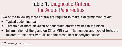
Diagnosing AP can be difficult because the signs and symptoms are similar to those of other medical conditions. The diagnosis is usually based upon a medical history, a physical examination, and the results of diagnostic tests (see TABLE 1).

Once a diagnosis of AP is made, additional tests are needed to determine the underlying cause. This ensures that the correct treatment is given to prevent pancreatitis from recurring.9
Imaging Tests: Imaging tests provide information about the structure of the pancreas, the ducts that drain the pancreas and gallbladder, and the surrounding tissues. Tests may include an x-ray of the abdomen (may show dilated loops of intestine or, rarely, one or more gallstones), x-ray of chest, CT scan (detecting inflammation of pancreas), or MRI of the abdomen.10
ERCP: ERCP is a procedure that can be used to remove stones from the bile duct if the pancreatitis is due to gallstones or other problems with the bile or pancreatic ducts.
Blood Test: No single blood test proves the diagnosis of AP, but certain tests suggest it. Blood levels of two enzymes produced by the pancreas, amylase and lipase, usually increase on the first day of the illness but return to normal in 3 to 7 days. If the person has had other flare-ups of pancreatitis, however, the levels of these enzymes may not increase because so much of the pancreas may have been destroyed that few cells are left to release the enzymes. The white blood cell count is usually increased.10
Ultrasound: An ultrasound may show gallstones in the gallbladder or common bile duct and may also detect swelling of the pancreas.
Other Techniques: If clinicians suspect the pancreas is infected, they may withdraw a sample of infected material from the pancreas by inserting a needle through the skin into the pancreas. Magnetic resonance cholangiopancreatography, a special MRI test, may also be done.
Treatment
The goals of treatment of AP are to alleviate pancreatic inflammation and to correct the underlying cause. Treatment usually requires hospitalization for at least a few days.
Mild Pancreatitis: Mild pancreatitis usually resolves with simple supportive care, which entails monitoring, drugs to control pain, and IV fluids. Patients may not be allowed to eat anything during the first few days if they have nausea or vomiting. Mild pancreatitis requires short-term hospitalization.11
Moderate-to-Severe Pancreatitis: Severe pancreatitis can lead to potentially life-threatening complications, including damage to the heart, lungs, and kidneys. Therefore, moderate-to-severe pancreatitis requires more extensive monitoring and supportive care. People with pancreatitis of this severity may be closely monitored in an intensive care unit where vital signs (pulse, blood pressure, and rate of breathing and urine production) can be monitored continuously. Blood samples are repeatedly drawn to monitor various components of the blood, including hematocrit, sugar (glucose) levels, electrolyte levels, white blood cell count, and amylase and lipase levels. Clinicians also give histamine2 blockers or proton pump inhibitors, which reduce or prevent the production of stomach acid.12
For patients who experience a drop in blood pressure or are in shock, blood volume is carefully maintained with IV fluids and heart function is closely monitored. Some people need supplemental oxygen, and the most seriously ill require a ventilator.
Most people with moderate-to-severe pancreatitis will not be able to eat in the early course of their illness. Instead, they may be fed through a tube placed through the nose or mouth into the small intestine (NG-tube) or through an IV line (TPN). IV fluids are given to help prevent dehydration. Patients can resume eating gradually once the pain resolves and bowel function returns to normal.
Acute pancreatitis is sometimes complicated by extensive damage and/or infection to the pancreatic tissue. In these cases, the damaged and/or infected tissue may be removed in a procedure referred to as a necrosectomy. Necrosectomy can be done as a minimally invasive procedure.12
Gallstone Pancreatitis: Gallstone pancreatitis recurs in 30% to 50% of people after an initial attack of pancreatitis. Surgical removal of the gallbladder (cholecystectomy) is often recommended during the same admission in mild cases to prevent a recurrence.
In elderly patients or those with serious medical problems, it may not be safe to remove the gallbladder. In this case, ERCP can be done to enlarge the bile duct opening. This allows stones from the gallbladder to pass, helping to prevent a recurrence of AP.11,12
Advances in diagnostic and therapeutic interventions have led to a decrease in mortality from AP, especially in those with severe, often necrotizing pancreatitis. Mortality in AP is usually due to systemic inflammatory response syndrome and organ failure in the first 2 weeks; after that period, it is usually due to sepsis and its complications.13
An infection is treated with antibiotics, and surgical removal of infected and dead tissue may be necessary. When pancreatitis is caused by a severe blunt or penetrating injury or uncontrolled biliary sepsis, surgeons perform surgery within the first several days to save the patient’s life.13
REFERENCES
1. Tenner S, Baillie J, Dewitt J, et al. American College of Gastroenterology guidelines: management of acute pancreatitis. Am J Gastroenterol. 2013;108(9):1400-1415.
2. Telem DA, Bowman K, Hwang J, et al. Selective management of patients with acute biliary pancreatitis. J Gastrointest Surg. 2009;13(12):2183-2188.
3. Whitcomb DC. Clinical practice. Acute pancreatitis. N Engl J Med. 2006;354(20):2142-2150.
4. Imrie CW. Prognostic indicators in acute pancreatitis. Can J Gastroenterol. 2003;17(5):325-328.
5. Kaurich T. Drug-induced acute pancreatitis. Proc Bayl Univ Med Cent. 2008;21(1):77-81.
6. Granger J, Remick D. Acute pancreatitis: models, markers, and mediators. Shock. 2005;24(suppl 1):45-51.
7. Haydock MD, Mittal A, van den Heever M, et al. National survey of fluid therapy in acute pancreatitis: current practice lacks a sound evidence base. World J Surg. 2013;3710:2428-2435.
8. Balthazar EJ, Ranson JH, Naidich DP, et al. Acute pancreatitis: prognostic value of CT. Radiology. 1985;156(3):767-772.
9. Ai X, Qian X, Pan W, et al. Ultrasound-guided percutaneous drainage may decrease the mortality of severe acute pancreatitis. J Gastroenterol. 2010;45(1):77-85.
10. Banks PA. Epidemiology, natural history, and predictors of disease outcome in acute and chronic pancreatitis. Gastrointest Endosc. 2002;56(suppl 6):S226-S230.
11. Morinville VD, Barmada MM, Lowe ME. Increasing incidence of acute pancreatitis at an American pediatric tertiary care center: is greater awareness among physicians responsible? Pancreas. 2010;39(1):5-8.
12. Balthazar EJ, Robinson DL, Megibow AJ, et al. Acute pancreatitis: value of CT in establishing prognosis. Radiology. 1990;174(2):331-336.
13. Maraví-Poma E, Gener J, Alvarez-Lerma F, et al. Early antibiotic treatment (prophylaxis) of septic complications in severe acute necrotizing pancreatitis: a prospective, randomized, multicenter study comparing two regimens with imipenem-cilastatin. Intensive Care Med. 2003;29(11):1974-1980.
To comment on this article, contact rdavidson@uspharmacist.com.


