US Pharm. 2014;39(9):HS-8-HS-12.
Cellulitis affects the skin and tissues beneath the skin. It is an infection that also involves the skin’s deeper layers, the dermis and subcutaneous tissue. It is very different from impetigo, which is a skin surface infection. The main bacteria responsible for cellulitis are Streptococcus and Staphylococcus organisms, the same ones that can cause impetigo. Methicillin-resistant Staphylococcus aureus (MRSA) can cause cellulitis. There have been many reports that certain other bacteria, such as Hemophilus influenzae and Pneumococcus and Clostridium species, have caused cellulitis as well.1
Although cellulitis may occur anywhere on the body, the lower leg is the most common site of infection (particularly the shinbone), followed by the arm and then the head and neck areas. Cellulitis can develop in the abdomen and chest areas as well. Obese people can develop cellulitis in the abdominal skin. Special types of cellulitis are sometimes designated by the location of the infection. Examples include periorbital cellulitis, buccal cellulitis, facial cellulitis, and perianal cellulitis. Orbital cellulitis results from a microbial infection with subsequent inflammation of the postseptal aspect of the eyelids.2 It is important to know that some people with poor leg circulation often develop scaly redness on the shins and ankles called stasis dermatitis, which is not a bacterially induced cellulitis.2
This article discusses several aspects of this disease, with emphasis on diagnosis and treatment, and reviews the guidelines of the Infectious Diseases Society of America (IDSA) on cellulitis.
Etiology
The most common bacteria that cause cellulitis are beta-hemolytic streptococci (groups A, B, C, G, and F). In addition, a form of rather superficial cellulitis caused by streptococcus is called erysipelas and is characterized by a spreading hot, bright red circumscribed area on the skin with a sharp, raised border. Erysipelas is more prevalent in young children. A strain of streptococcal bacteria can sometimes rapidly destroy tissues beneath the skin.3
There is a growing incidence of community-acquired infections due to MRSA, a particularly dangerous form of staphylococcal bacteria that is resistant to many antibiotics and is therefore more difficult to treat.
Many other bacteria can cause cellulitis. In young children, H influenzae (H flu) bacteria can cause cellulitis, especially on the face and arms. Cellulitis from a dog or cat bite or scratch may be caused by the Pasteurella multocida bacterium, which has a very short incubation period of only 4 to 24 hours. Cellulitis can also be caused by Aeromonas hydrophila, Vibrio vulnificus, and other bacteria after exposure to freshwater or saltwater. Pseudomonas aeruginosa is another type of bacterium that can cause cellulitis, typically after a puncture wound.3
Signs and Symptoms
There are four signs and symptoms associated with cellulitis: erythema, pain, swelling, and warmth. The categories are as follows.
Uncomplicated cases of cellulitis: In these cases, the skin infection is without underlying drainage, penetrating trauma, or abscess and is most likely caused by a streptococcus or S aureus. There is limited area of involvement with minimal pain. This category includes conditions with no systemic signs of illness (e.g., fever, altered mental status, tachypnea, tachycardia, hypotension) and no risk factors for serious illness (e.g., extremes of age, general debility, immunocompromised health).4
Severe and complicated cases of cellulitis: These cases involve malaise, chills, fever, and toxicity; lymphangitic spread (red lines streaking away from the area of infection); circumferential cellulitis; and pain disproportionate to examination findings.
Emergent surgical evaluation: These cases include violaceous bullae, cutaneous hemorrhage, skin sloughing, skin anesthesia, rapid progression, and gas in the tissue.4
Risk Factors
Often, cellulitis develops in the area of a break in the skin, such as a cut, small puncture wound, or insect bite. In some cases, cellulitis develops due to microscopic cracks in the skin that are inflamed or irritated. It can also occur around surgical wounds.5
Cellulitis can develop where there is no skin break at all, such as a chronic leg swelling. Athlete’s foot or impetigo can also predispose a person to develop cellulitis. Other diseases conditions such as eczema and psoriasis or skin damage caused by radiation therapy can lead to cellulitis.
People with diabetes or those receiving chemotherapy or drugs that suppress the immune system are particularly prone to developing cellulitis. Conditions that reduce the circulation of blood in the veins or reduce circulation of the lymphatic fluid also increase the risk of developing cellulitis.
Tattoos usually are not considered risky procedures (an estimated 45 million people in the United States have at least one), but they do entail risk for infection with both typical bacterial pathogens and less common organisms. If infected, patients first complain of pruritic pustules in newly tattooed skin.6
Tattoo infections are widely underreported, and nontuberculous mycobacterial infections may be far more common than it is believed. This diagnosis is unlikely to be made without mycobacterial cultures of skin-biopsy specimens, and in many cases drug resistance is very common among these organisms.6
Cellulitis is not contagious because it is an infection of the skin’s deeper layers (the dermis and subcutaneous tissue), and the skin’s top layer (the epidermis) provides a cover over the infection. In this regard, cellulitis is different from impetigo, in which there is a very superficial skin infection that can be contagious.
Diagnosis
It is important first to establish that the inflammation is due to an infection. Past medical history and a physical examination can provide more information. Elevated white blood cell count and a culture of bacteria may also be of high value in diagnosis. However, in many cases of cellulitis, the concentration of bacteria may be low and cultures may fail to demonstrate the causative organism.4
When it is difficult or impossible to distinguish whether or not the inflammation is due to an infection, clinicians treat the patient with antibiotics just to be sure. If the condition is not resolved, the inflammation may not be due to an infection, and different methods dealing with types of inflammation may be applied. For example, if the inflammation is thought to be due to an autoimmune disorder, treatment with corticosteroids may be warranted.5
First, antibiotics, such as penicillin derivatives or other types of antibiotics, are used to treat cellulitis. If the bacterium turns out to be resistant to the chosen antibiotics or patients allergic to penicillin, other appropriate antibiotics can be substituted. In some cases, oral antibiotics may not always provide sufficient penetration of the inflamed tissues to be effective, and in these instances antibiotics will be administered in a hospital setting or at home.
The method of treatment is based on many factors, including the location and extent of the infection, the type of bacterium causing the infection, and the overall health status of the patient
Cellulitis can be prevented by proper hygiene, treating chronic swelling of tissues (edema), and care of wounds. It is preventable in a healthy person with a healthy immune system, but in people with predisposing conditions and/or weakened immune systems, cellulitis may not always be avoidable.2
Blood Tests and Culture
The following blood tests are recommended for patients with soft-tissue infection who have signs and symptoms of systemic toxicity: CBC with differential; levels of creatinine, bicarbonate, creatine phosphokinase, and C-reactive protein; and blood cultures.3 Blood cultures should be done in moderate-to-severe disease (e.g., cellulitis with complicating lymphedema); in cellulitis of specific anatomical sites (e.g., facial and especially ocular areas); and in patients with a history of contact with potentially contaminated water. Additional tests that may be warranted include mycologic investigation if recurrent episodes of cellulitis are suspected to be secondary to tinea pedis or onychomycosis, and serum creatinine levels to help assess baseline renal function and guide antimicrobial dosing.
Imaging Studies
If necrotizing fasciitis is a concern, CT imaging is typically used to examine stable patients. Ultrasonography may play a role in the detection of occult abscess and the direction of medical care, while ultrasonographic-guided aspiration of pus can shorten hospital stay and fever duration in children with cellulitis. If there is a strong clinical suspicion of necrotizing fasciitis, surgical consultation should be initiated without delay.
Aspiration and Biopsy
Needle aspiration should be performed only in selected patients or in unusual cases, such as in patients who have diabetes, are immunocompromised, are neutropenic, are not responding to empirical therapy, or have a history of animal bites. Gram stain and culture following incision and drainage of an abscess yield positive results in more than 90% of cases. Skin biopsy is not routine but may be performed in an attempt to rule out a noninfectious entity.7
Cellulitis Management and Treatment
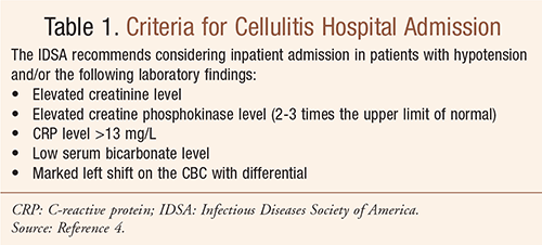
Cellulitis is a treatable condition, but antibiotics are necessary to eradicate the infection and prevent its spread. Most cellulitis can be effectively treated with oral antibiotics at home. Hospitalization and IV antibiotics are sometimes required if oral antibiotics are not effective (see TABLE 1). If not properly treated, cellulitis can occasionally spread to the bloodstream and cause sepsis, a serious bacterial infection.5 Antibiotic regimens to treat cellulitis are effective in more than 90% of patients. Regardless of the pathogen, all but the smallest of abscesses require drainage for resolution. If the abscess is relatively isolated, drainage only—without antibiotics—may suffice.5,8

In cases of cellulitis without draining wounds or abscess, streptococci continue to be the likely etiology, and beta-lactam antibiotics are appropriate therapy. In mild cases of cellulitis treated on an outpatient basis, dicloxacillin, amoxicillin, or cephalexin may be used. In patients who are allergic to penicillin, clindamycin or a macrolide (clarithromycin or azithromycin) may be appropriate. An initial dose of a parenteral antibiotic with a long half-life (e.g., ceftriaxone) followed by an oral agent may also be called for.5,8
In cases of recurrent disease, most often due to Streptococcus species (usually related to venous or lymphatic obstruction), penicillin G, amoxicillin, or erythromycin may be effective. If tinea pedis is suspected to be the predisposing cause, treatment with topical or systemic antifungals is necessary.5,8
Patients with severe cellulitis require parenteral therapy with broad gram-positive, gram-negative, and anaerobic antibiotics, including cefazolin, cefuroxime, ceftriaxone, nafcillin, or oxacillin for presumed staphylococcal or streptococcal infection. Until culture and sensitivity information becomes available, these medications are also warranted as coverage for MRSA for severe cellulitis apparently related to an abscess. In patients who are allergic to penicillin, vancomycin and clindamycin are appropriate.5,8
For cellulitis involving wounds in water, recommended antibiotic regimens vary with the type of water involved. For freshwater involvement, a third- or fourth-generation cephalosporin (e.g., ceftazidime or cefepime) or a fluoroquinolone (e.g., ciprofloxacin or levofloxacin) is used. In cases of saltwater exposure, doxycycline and ceftazidime or a fluoroquinolone (e.g., levofloxacin) is called for. Lack of response to an appropriate antibiotic regimen should raise suspicion for Mycobacterium marinum infection and suggests wound biopsy for mycobacterial stains and culture.5,8
REFERENCES
1. Pasternack MS, Swartz MN. Cellulitis, necrotizing fasciitis and subcutaneous tissue infections. In: Mandell GL, Bennett JE, Dolin R, eds. Mandell, Douglas, and Bennett’s Principles and Practice of Infectious Diseases. Vol. 1. 7th ed. Philadelphia, PA: Churchill Livingstone Elsevier; 2010:1289-1312.
2. Parvizi N, Choudhury N, Singh A. Complicated periorbital cellulitis: case report and literature review. J Laryngol Otol. 2012;126(1):94-96.
3. Heagerty AH. Cellulitis and erysipelas. In: Lebwohl MG, Coulson I, Heymann WR, Berth-Jones J, eds. Treatment of Skin Disease: Comprehensive Therapeutic Strategies. 3rd ed. Edinburgh, Scotland: Saunders Elsevier; 2010:132-134.
4. Freedberg IM, Eisen AZ, Wolff K, et al. Soft-tissue infections: erysipelas, cellulitis, gangrenous cellulitis, and myonecrosis. In: Wolff K, et al, eds. Fitzpatrick’s Dermatology in General Medicine. 7th ed. New York, NY: McGraw-Hill Medical; 2008;7:1720-1731.
5. Habif TP. Cellulitis and erysipelas section of bacterial infections. In: Clinical Dermatology: A Color Guide to Diagnosis and Therapy. 5th ed. Philadelphia, PA: Mosby; 2010:342-350.
6. Falsey RR, Kinzer MH, Hurst S, et al. Cutaneous inoculation of nontuberculous mycobacteria during professional tattooing: a case series and epidemiologic study. Clin Infect Dis. 2013;57-60.
7. Howe L, Jones N. Guidelines for the management of periorbital cellulitis/abscess. Clin Otolaryngol Allied Sci. 2004;29(6):725-728.
8. Lexi-Comp Online Drug Information Database, 2012.
To comment on this article, contact
rdvaidson@uspharmacist.com.


